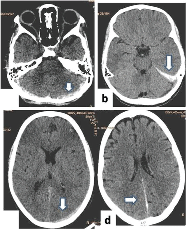
Figure 3:
NCCT scans axial images of different patient shows different location of SDH, (a) shows SDH along bilateral cerebellar hemispheres, (b) SDH along tentorium on left side, (c) SDH along left occipital lobe, (d) SDH along posterior falx.